


Most people book an eye test because their vision feels blurry, their glasses no longer feel right, or they are struggling with screens, reading or night driving. But some of the most important parts of an eye examination are not about how clearly you can read letters on a chart. They are about the health of the retina the light-sensitive layer at the back of the eye that plays a vital role in how you see.
The challenge is that retinal changes can develop quietly. You may not feel pain. You may not notice symptoms straight away. In some cases, by the time vision changes become obvious, the condition may already need more urgent attention. This is why advanced retinal imaging has become such an important part of modern eye care.
The Optos Daytona helps optometrists capture a much wider view of the retina in a quick, comfortable, and highly detailed way. For patients, it means a clearer understanding of what is happening inside the eye. For eye care professionals, it provides a valuable image that can be reviewed, compared and monitored over time.
What Is the Optos Daytona?
The Optos Daytona is an advanced retinal imaging device used to take ultra-widefield images of the back of the eye. Instead of only capturing a small central section of the retina, it gives the optometrist a much broader view, including areas toward the outer edges of the retina that can be harder to examine with standard imaging.
In simple terms, it works like a high-quality panoramic photograph of your retina. During the scan, you look into the device, follow positioning guidance and the image is captured in a fraction of a second. There is no contact with the eye and the process is usually straightforward for both adults and children.
Why the Wider View Matters
A standard retinal photograph may show the central area of the retina clearly, which is useful for checking the macula, optic nerve, and blood vessels. However, some signs of retinal problems can appear further out in the peripheral retina. A wider image gives the optometrist more information in one scan, helping them assess your eye health more thoroughly.
This does not mean every patient has a serious condition hiding in the background. It means your optometrist has a better view, better documentation and a stronger basis for deciding whether your eyes look healthy or need closer attention.
How the Optos Daytona Works During an Eye Test
The scan is designed to be quick and comfortable. You place your face near the device, look at a target, and the system captures an ultra-wide image of the retina. The optometrist can then review the image on screen and explain what they can see.
A More Visual Eye Care Experience
One of the most useful parts of Optos Daytona imaging is that patients can often see their own retinal image. This turns the eye test into a clearer more practical conversation.
For example, instead of simply being told that a blood vessel needs monitoring, a patient can be shown the exact area on the retinal image. This makes advice easier to understand and can help patients take follow-up recommendations more seriously.
A Digital Record for Future Comparison
Because the image is digital, it can be stored as part of your eye health record. This is especially valuable for monitoring small changes over time. If you return for another eye test in the future, your optometrist can compare the latest image with previous scans.
That comparison can be helpful when checking whether a mark, lesion, blood vessel change or retinal feature has remained stable or changed. In eye care, change over time often matters just as much as what is visible on the day.
What Can Optos Daytona Help Your Optometrist See?
The Optos Daytona supports a more detailed look at the retina and surrounding structures. It is commonly used as part of a wider eye health assessment and can help with the observation of several areas of concern.
It may help your optometrist review signs linked to:
- Diabetic eye changes, including changes in retinal blood vessels
- Macular changes that may affect central vision
- Retinal tears, thinning or suspicious peripheral changes
- Choroidal lesions or pigmented marks at the back of the eye
- Optic nerve appearance, which may be relevant in glaucoma monitoring
- General retinal health in patients with family history or risk factors
The key point is that Optos Daytona imaging supports assessment. It does not replace professional judgement and it does not mean every finding is a diagnosis. If something needs further investigation, your optometrist may recommend additional imaging, dilation, OCT scanning, monitoring or referral to an eye specialist.
Why It Is Useful for People With No Symptoms
Many patients assume that if they can see well, their eyes must be healthy. Unfortunately, the retina does not always follow that rule. Some retinal and vascular changes can begin without obvious symptoms.
This is one reason regular eye tests are important, even when your vision feels normal. Advanced imaging adds another layer of information by helping your optometrist examine and document the back of the eye more clearly.
Everyday Examples Where Retinal Imaging Helps
Consider someone with diabetes who has no noticeable vision problems. Retinal imaging may help the optometrist look for early blood vessel changes and decide whether routine monitoring or further care is needed.
Or think about someone with a strong family history of eye disease. Even if their prescription has barely changed, a retinal image provides a useful baseline for future comparison.
For a patient who notices new floaters, flashes, or shadows a wide view of the retina may help the optometrist assess whether the symptoms need urgent attention.
Optos Daytona vs Traditional Retinal Photography
Traditional retinal photography still has value, especially for viewing the central retina. However, its field of view is more limited. The Optos Daytona is designed to capture a much wider retinal image in one scan, giving the optometrist more peripheral detail.
The Difference in Practical Terms
With a narrower image, the optometrist may only see the central part of the retina. With an ultra-widefield image, they can view much more of the retinal surface at once. This wider perspective can be especially useful when checking areas that are not always easy to document with standard imaging.
It also makes patient education easier. A wide retinal image can show the bigger picture, helping patients understand why their eye health is being monitored and what the optometrist is looking for.
Is Optos Daytona Imaging Comfortable?
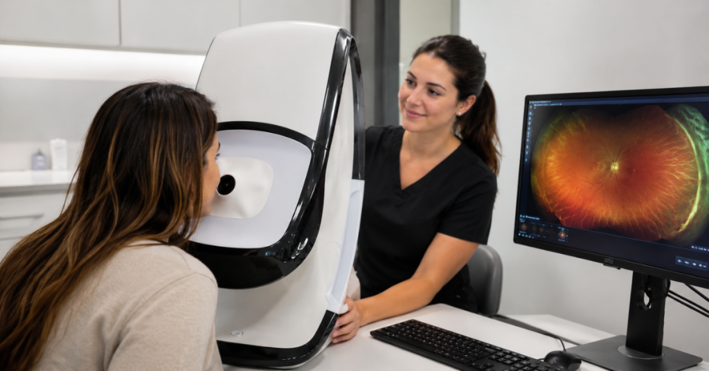
For most patients, the scan is quick, non-invasive and comfortable. There is no physical contact with the eye. You may notice a brief flash or light during image capture, but the experience is usually over very quickly.
Helpful for Nervous Patients and Children
Some people feel anxious about eye tests, especially if they dislike drops, bright lights or instruments close to the eye. The Optos Daytona can make retinal imaging feel more approachable because the scan is fast and simple.
For children, older patients or people who struggle to sit still for long examinations, this can make a noticeable difference. A smoother experience often means better-quality images and less stress during the appointment.
Does Optos Daytona Replace Dilation?
Optos Daytona imaging is a powerful tool, but it does not completely replace every other method of examining the eye. In some cases, your optometrist may still recommend dilation drops to get a different or more detailed view.
Dilation may be needed when symptoms are present, when the view is unclear or when a specific area requires closer examination. Similarly, OCT scans may be recommended when the optometrist needs a layered cross-sectional view of the retina.
The best approach is not about choosing one technology over another. It is about using the right tools for the patient’s eye health needs.
Who May Benefit Most From Optos Daytona Imaging?
Optos Daytona imaging can be useful for many patients, but it is especially helpful when a wider retinal view or long-term monitoring is valuable.
You may particularly benefit if you:
- Have diabetes, high blood pressure or other vascular risk factors
- Have a family history of glaucoma, macular degeneration or retinal disease
- Notice flashes, floaters, shadows or sudden changes in vision
- Have previously been told you have a retinal mark or lesion
- Want a detailed baseline image for future comparison
- Prefer a quick, non-contact retinal imaging experience
This does not mean the scan is only for people with known problems. Many patients choose advanced retinal imaging because they want a more complete understanding of their eye health before symptoms appear.
Why Optos Daytona Matters for Better Patient Care
The biggest value of Optos Daytona imaging is not just the technology itself. It is what the image allows your optometrist to do with more confidence.
A clear, wide retinal image can support better conversations, better monitoring and better decision-making. It helps move eye care from a simple “your prescription has changed” appointment to a fuller assessment of long-term visual health.
For practices such as City Specs, this type of imaging also improves the patient experience. People can see what the optometrist is discussing, ask better questions and leave with a clearer understanding of their eye health.
What to Expect After the Scan
After the scan, your optometrist will review the image and explain any relevant findings. If everything looks healthy, the image can still be useful as a record for future visits. If something needs monitoring, your optometrist can explain the next steps.
Possible next steps may include:
- Routine monitoring at your next eye test
- A follow-up scan after a set period
- Additional imaging such as OCT
- Dilated examination for a closer view
- Referral to a hospital eye service or specialist if needed
The important thing is that the image gives both you and your optometrist more information to work with.
Conclusion
The Optos Daytona gives optometrists a clearer and wider view of the retina, helping them assess areas that may be difficult to see with standard imaging alone. For patients, it makes eye care more visual, more understandable, and more proactive.
Its value is especially strong in early observation, long-term monitoring and patient education. A single image can provide a baseline that supports future comparisons, helping your optometrist spot changes that may otherwise be harder to explain or track.
As eye care continues to become more personalised, tools like the Optos Daytona will play an important role in helping people look after their sight before problems become obvious. It is not a replacement for every clinical test, but it is a powerful addition to a modern eye examination giving both patient and optometrist a clearer picture of retinal health.
FAQs
What is Optos Daytona used for?
Optos Daytona is used to capture ultra-widefield images of the retina, helping optometrists examine and monitor the back of the eye more clearly.
Is Optos Daytona imaging painful?
No. The scan is non-contact, quick and usually very comfortable. Most patients only notice a brief flash of light.
Does Optos Daytona replace a normal eye test?
No. It supports the eye test by giving a wider retinal image, but your optometrist will still carry out other checks as needed.
Can Optos Daytona detect eye disease?
It can help optometrists identify signs that may need monitoring or further investigation, but diagnosis depends on a full professional assessment.
Do I still need dilation if I have Optos Daytona imaging?
Sometimes, yes. Your optometrist may still recommend dilation if they need a closer or different view of the retina.


